Central Vestibular System: How to Differentiate Central vs. Peripheral Dysfunction
Don’t let complex vestibular cases derail care. Gain practical insights to help you distinguish central from peripheral dysfunction and tailor your interventions.
June 18, 2025
9 min. read
The central vestibular system plays a critical role in how we interpret movement, maintain balance, and stabilize gaze. Yet its dysfunction often presents alongside or is mistaken for peripheral vestibular issues. Clinicians regularly encounter patients with dizziness, imbalance, and visual disturbances, but distinguishing whether these symptoms stem from central or peripheral vestibular pathways is not always straightforward.
Accurate differentiation is essential for guiding assessment, clinical reasoning, and treatment planning. In this article, we’ll examine the key distinctions between central vs. peripheral vestibular system dysfunction and outline clinical strategies to help therapists make more informed decisions during vestibular rehabilitation.
The vestibular system: Structure and function
The vestibular system is at the core of every well-executed head turn, upright posture, or smooth visual focus. This intricate network is responsible for sensing motion, orienting the body in space, and maintaining gaze stability. It’s not just anatomy. It allows patients to walk confidently, look around without dizziness, and feel grounded in their environment.
Understanding the vestibular system’s architecture is foundational to identifying where dysfunction may occur. Clinically, we divide this system into peripheral and central components, each with distinct structures and functions that guide how we assess and intervene.
Peripheral vestibular system
Vestibular end organs:
Three semicircular canals (detecting angular acceleration)
Two otolith organs (utricle and saccule, detecting linear acceleration and gravity)
Vestibular portion of cranial nerve VIII, which transmits signals to the brainstem
Central vestibular system
Vestibular nuclei (lateral, medial, superior, inferior) at the pontomedullary junction
Vestibulocerebellum (flocculonodular lobe and vermis)
Vestibulo-ocular pathways (modulating gaze stability via the vestibulo-ocular reflex)
Vestibulospinal pathways (contributing to postural control)
Vestibular areas in the cortex (parietal-insular vestibular cortex, thalamus), supporting spatial orientation, motion perception, and multisensory integration
Vestibulo-autonomic pathways (influencing autonomic responses and emotional regulation)
Clinical presentation: Central vs. peripheral dysfunction
Dizziness is a complex and multifaceted symptom. Few assessments are as nuanced as determining whether it stems from central or peripheral vestibular dysfunction. It’s a familiar puzzle: a patient describes vertigo, visual instability, or unsteadiness, and you’re tasked with pinpointing the source. While symptoms often overlap, their origins carry very different implications for treatment and recovery.
Recognizing the differences between central and peripheral patterns is key to diagnosing and choosing the proper treatment. Here’s a breakdown of hallmark presentations to support your clinical decision-making.
Peripheral vestibular dysfunction
Peripheral vestibular dysfunction is often more familiar and straightforward to assess. When the vestibular end organs or nerves are impaired, symptoms tend to follow recognizable patterns and respond well to targeted rehabilitation.
Patients may report spinning or positional vertigo, particularly with head movements. Fortunately, many peripheral disorders (such as BPPV, vestibular neuritis, and Ménière’s disease) respond well to interventions like canalith repositioning or gaze stability training.
Here are the hallmark features you’ll often observe:
Vertigo triggered by head movements
True vertigo (sensation of spinning)
Nystagmus suppressed by visual fixation
Possible hearing loss (with cochlear involvement)
Central vestibular dysfunction
Central vestibular dysfunction presents a greater diagnostic challenge, often producing more complex and persistent symptom patterns.
When the vestibular nuclei, cerebellum, or cortical pathways are disrupted, patients may experience more than just gaze instability and motion sensitivity. Symptoms can also include visual motion intolerance, an impaired sense of midline or spatial orientation, and even autonomic or emotional responses that complicate recovery.
Unlike peripheral conditions, central dysfunction is less likely to resolve fully with standard interventions, requiring a more nuanced approach that addresses sensory, motor, and emotional dimensions.
These are the hallmark features you may observe:
Impaired midline orientation
Direction-changing or multidirectional nystagmus not suppressed by fixation
Impaired smooth pursuit and saccades
Imbalance and postural instability, potentially out of proportion to the sensation of dizziness
Visual motion intolerance and sensitivity in complex environments
Pronounced emotional responses (anxiety, panic) driven by autonomic and limbic system connections
Clinicians should remain mindful that central and peripheral dysfunction can co-occur, particularly in older adults and those with complex neurological presentations.
Vestibular pathways and clinical assessment
What makes the vestibular system so powerful and diagnostically rich is how its functions are organized into distinct but overlapping neural pathways, each contributing to the patient’s functional performance and symptom profile.
When symptoms don’t fit a clean peripheral pattern, it’s often because central modulation of reflexes or multisensory integration is disrupted. Here are the four main vestibular pathways clinicians should keep in mind during assessment:
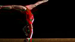
Vestibulo-ocular reflex (VOR): Stabilizes vision by coordinating compensatory eye movements during head motion. The cerebellum fine-tunes this reflex, especially when the patient is tracking a moving target (as illustrated in the soccer example from the course).
Vestibulospinal reflex (VSR): Supports postural control through reflexive activation of trunk and limb musculature. This system contributes to both short-latency reflexes and long-latency postural adjustments, which are critical when assessing imbalance and fall risk.
Vestibulo-autonomic pathways: Regulate blood pressure, arousal, and stress responses during postural transitions. Clinically, dysfunction here often manifests as lightheadedness, nausea, or disproportionate anxiety during vestibular stimulation.
Cortical vestibular pathways: Support spatial orientation, self-motion perception, and multisensory integration. Impairments in these pathways can present as visual motion sensitivity, poor vertical alignment, or disorientation in visually complex environments, symptoms commonly seen in central vestibular dysfunction.
When approaching vestibular assessment, mapping symptoms to these pathways provides critical diagnostic clues. A thorough evaluation should include a detailed oculomotor exam, sensory organization testing, and static and dynamic postural control assessments. Together, these offer a direct window into how well these pathways, particularly those influenced by the central vestibular system, function and integrate with other sensory systems.
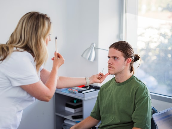
The role of the oculomotor exam
The oculomotor test is a critical part of assessing the central vestibular system. Key elements to determine include:
Extraocular movements (ROM): Assess the range and conjugacy of eye movements. Limitations or disconjugate gaze suggest central involvement or cranial nerve impairment.
Smooth pursuit: Ability to smoothly track a moving target. Saccadic intrusions indicate central dysfunction.
Saccades: Rapid eye movements between targets. Hypometric, hypermetric, or delayed saccades are indicative of central lesions.
VOR suppression (cancellation): Ability to suppress the VOR when tracking a moving target with eyes and head moving together. Cerebellar dysfunction often impairs this ability.
Convergence: Assess the near point of convergence (normal is 5 to 7 centimeters). Impairments can reflect either central or peripheral involvement.1
Dynamic visual acuity: Compare static and dynamic visual acuity. A decline of three or more lines is abnormal but not specific to central or peripheral dysfunction.2
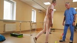
Postural responses and central integration
The vestibular system’s influence on postural control extends well beyond simple reflexes. Automatic postural responses are layered, time-sensitive, and deeply modulated by both subcortical and cortical systems.
Understanding how these responses are organized helps explain why some patients struggle with anticipatory control or postural adjustments in complex environments.
Automatic postural responses
Short-latency (SL): Rapid reflexive responses (e.g., ankle dorsiflexion after perturbation) driven primarily by spinal and peripheral mechanisms.
Medium-latency (ML): Responses modulated by the vestibular nuclei and basal ganglia are important for coordinated postural corrections.
Long-latency (LL): Cortically mediated responses that allow for anticipatory adjustments and feedforward control, especially during voluntary movement.
Cerebellar contributions
The vestibulocerebellum plays a central role in refining both the vestibulo-ocular reflex and the vestibulospinal reflex by monitoring and adjusting vestibular reflexes in real time and enabling cancellation of reflexes when appropriate—for example, by suppressing the VOR to follow a moving object or modulating postural responses during voluntary movement.
When damaged, cerebellar dysfunction can present as gaze instability, nystagmus, ataxia, and imbalance, which are key signs that central pathways are compromised.
Emotional and autonomic pathways
Vestibular dysfunction often engages autonomic and emotional systems, contributing to symptom severity and functional limitations:
Locus coeruleus: Triggers stress and panic responses.
Nucleus of the solitary tract: Generates nausea via vagal pathways.
Area postrema: Mediates vomiting.
Amygdala: Creates strong emotional memories of vestibular crises.
This helps explain why some patients develop anxiety, avoidance behaviors, or heightened emotional reactivity, even after the original vestibular insult has resolved. These reactions are not voluntary or purely psychological—they are hardwired neurophysiological responses.
As therapists, recognizing and validating these responses is critical. Addressing them through graded exposure, education, and supportive rehab strategies can significantly improve patient outcomes and engagement.
Clinical implications and treatment
By identifying which vestibular pathways are involved, clinicians can design more effective, individualized treatment plans. Targeted interventions should promote compensation and address the complex interplay between motor, sensory, autonomic, and emotional systems.
Peripheral dysfunction
Employ canalith repositioning techniques to resolve positional vertigo in BPPV.
Use gaze stability and habituation exercises to promote central compensation and restore functional head and eye movements.
Central dysfunction
Focus on postural control retraining with graded exposure to progressively challenge balance and sensory integration.
Implement visual motion desensitization strategies to improve tolerance of visually complex environments.
Address emotional and cognitive responses through graded exposure, patient education, reassurance, and collaboration with behavioral health specialists as needed.
Applying central and peripheral vestibular insights to practice
Differentiating between central and peripheral vestibular dysfunction is fundamental to providing targeted, effective care. By combining knowledge of vestibular pathways with thoughtful clinical assessment and by addressing the emotional and autonomic dimensions that often accompany dysfunction, you can better support patients in regaining stability, function, and confidence.
For a more in-depth exploration of these pathways and practical assessment techniques, you can learn more through our Medbridge course, Anatomy and Physiology of the Central Vestibular System. The course provides a comprehensive look at how the central vestibular system integrates sensory information to support balance, gaze stability, and postural control, offering you actionable strategies to enhance assessment and intervention for individuals with central vestibular dysfunction.
References
Scheiman, M., Gallaway, M., Frantz, K. A., Peters, R. J., Hatch, S., Cuff, M., & Mitchell, G. L. (2003). Nearpoint of convergence: test procedure, target selection, and normative data. Optometry and vision science : official publication of the American Academy of Optometry, 80(3), 214–225. http://www.ncbi.nlm.nih.gov/pubmed/12637833
Longridge, N. S., & Mallinson, A. I. (1987). The dynamic illegible E-test. A technique for assessing the vestibulo-ocular reflex. Acta oto-laryngologica, 103(3-4), 273–279. https://pubmed.ncbi.nlm.nih.gov/3577760/
Below, watch Laura Morris discuss peripheral vs. central vestibular system in this brief clip from her and Jeffrey Hoder's Medbridge course "Anatomy and Physiology of the Central Vestibular System."

Meet the Authors


Subscribe to Our Newsletter
Related Posts
July 30, 2021
The Mighty Vestibular System: Balance and Beyond
By Medbridge

December 15, 2015
Concussion: What Acute Sign Is Most Important?
By Anne Mucha and Susan Whitney
April 9, 2024
9 Effective Exercises for Vestibular Hypofunction
By Morgan Kriz

September 8, 2019
An Essential Exercise for Head Motion Induced Oscillopsia
By Jeff Walter